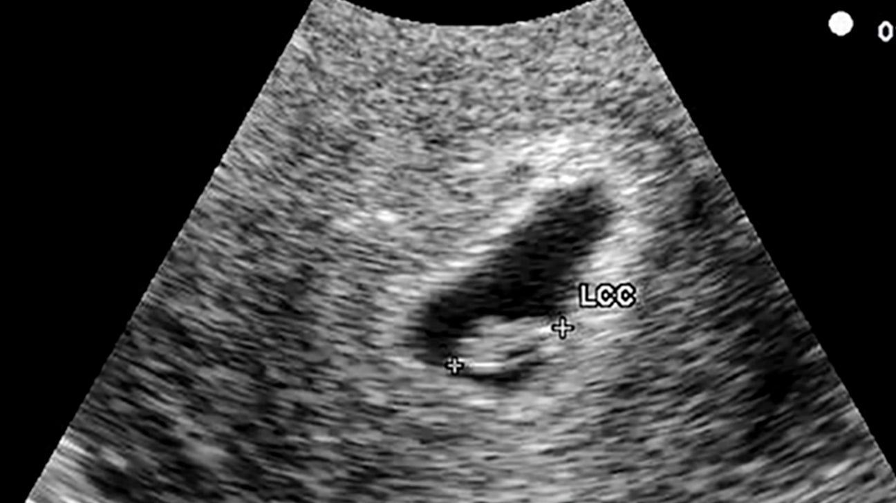
Ultrasound of 6 Week Pregnancy: A Comprehensive Guide
An ultrasound at 6 weeks of pregnancy offers an early glimpse into the development of your baby. This non-invasive imaging technique uses sound waves to create images of the uterus and its contents. Here’s a comprehensive guide to what you can expect during a 6-week ultrasound:
What to Expect During the Ultrasound
- Preparation: Typically, no special preparation is required for a 6-week ultrasound. However, you may be asked to drink plenty of fluids beforehand to fill your bladder, which helps improve the visibility of the uterus.
- Procedure: The ultrasound is performed transvaginally, meaning a small probe is inserted into the vagina to obtain images of the uterus. The procedure usually takes around 15-20 minutes.
- Positioning: You will lie on your back with your knees bent and your feet in stirrups. The probe will be gently inserted into the vagina and moved around to capture images of the uterus.
What the Ultrasound Can Show
At 6 weeks of pregnancy, the ultrasound may reveal:
- Gestational sac: A small, fluid-filled sac that contains the developing embryo.
- Yolk sac: A small, dark circle within the gestational sac that provides nutrients to the embryo.
- Embryo: A tiny, bean-shaped structure that will eventually develop into your baby.
- Heart activity: The ultrasound may be able to detect the embryo’s heartbeat, which appears as a flickering light on the screen.
Interpreting the Results
The ultrasound findings will be interpreted by your healthcare provider. They will assess the following:
- Size and location of the gestational sac: The size of the gestational sac should correspond to the expected gestational age.
- Presence of the yolk sac: The yolk sac should be visible and located within the gestational sac.
- Embryo size and development: The embryo should be visible and its size should be appropriate for the gestational age.
- Heart activity: The presence of a heartbeat is a positive sign that the embryo is developing normally.
What if the Ultrasound is Abnormal?
In some cases, the ultrasound may reveal abnormalities, such as:
- Empty gestational sac: If the gestational sac is empty, it may indicate a miscarriage or an ectopic pregnancy.
- Abnormal yolk sac: An enlarged or misshapen yolk sac may be associated with certain genetic abnormalities.
- No embryo visible: If the embryo is not visible at 6 weeks, it may be too early to detect or could indicate a miscarriage.
- Abnormal heart activity: A slow or irregular heartbeat may be a sign of developmental problems.
Follow-Up Care
Depending on the ultrasound findings, your healthcare provider may recommend follow-up care, such as:
- Repeat ultrasound: If the ultrasound is inconclusive or abnormal, a repeat ultrasound may be scheduled to confirm the findings.
- Blood tests: Blood tests can check for pregnancy hormones and other markers to assess the health of the pregnancy.
- Genetic testing: Genetic testing may be recommended if there are concerns about the embryo’s development.
Emotional Impact of the Ultrasound
Seeing your baby for the first time on an ultrasound can be an emotional experience. It can evoke feelings of joy, excitement, and anticipation. However, it’s important to remember that the ultrasound is just a snapshot in time and does not guarantee a healthy pregnancy.
Conclusion
An ultrasound at 6 weeks of pregnancy provides valuable information about the early development of your baby. While it can be a reassuring experience, it’s important to interpret the results in context and follow your healthcare provider’s recommendations for follow-up care. The ultrasound is a valuable tool that can help you and your healthcare provider monitor the progress of your pregnancy and ensure the well-being of your baby.