Ultrasound Pictures of Pregnancy at 8 Weeks
An ultrasound is a medical imaging technique that uses high-frequency sound waves to produce images of the inside of the body. During pregnancy, an ultrasound can be used to monitor the development of the fetus and to assess the health of the mother.
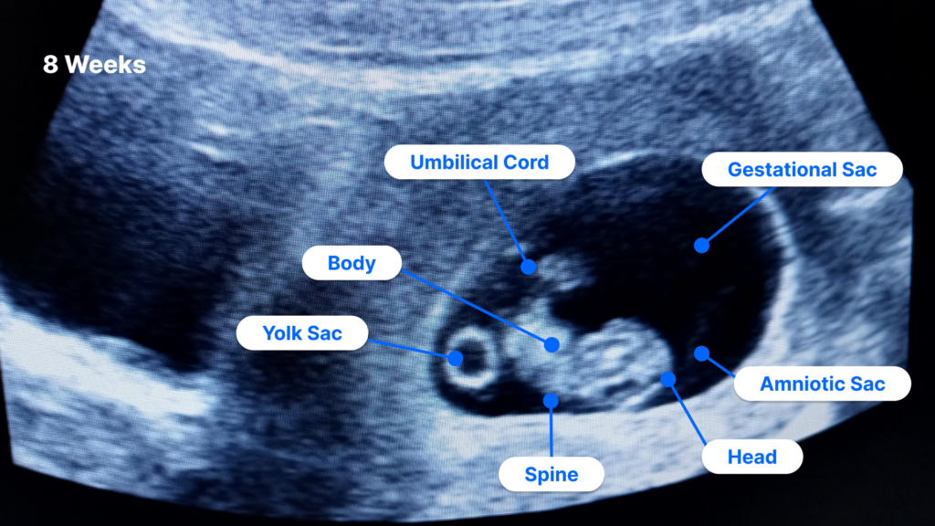
At 8 weeks of pregnancy, the fetus is about the size of a grape. The head is large in proportion to the body, and the facial features are beginning to develop. The arms and legs are also starting to form, and the heart is beating regularly.
An ultrasound at 8 weeks can provide valuable information about the pregnancy. The doctor can use the ultrasound to:
- Confirm the pregnancy
- Determine the gestational age of the fetus
- Check the fetal heart rate
- Measure the fetus’s growth
- Look for any abnormalities
An ultrasound at 8 weeks can also be used to perform a chorionic villus sampling (CVS). CVS is a prenatal test that can be used to diagnose certain genetic disorders.
What to Expect During an Ultrasound at 8 Weeks
An ultrasound at 8 weeks is typically performed transvaginally. This means that the ultrasound probe is inserted into the vagina. The probe emits sound waves that bounce off the fetus and the surrounding tissues. The sound waves are then converted into images that can be viewed on a monitor.
An ultrasound at 8 weeks is usually painless. However, some women may experience some discomfort when the probe is inserted into the vagina.
The ultrasound will typically take about 15-20 minutes. The doctor will explain the results of the ultrasound to you and answer any questions you may have.
What the Ultrasound Pictures Show
The ultrasound pictures at 8 weeks will show the fetus in the uterus. The head will be large in proportion to the body, and the facial features will be beginning to develop. The arms and legs will also be starting to form, and the heart will be beating regularly.
The doctor will also be able to measure the fetus’s growth. The fetus’s length will be measured from the crown of the head to the rump. The doctor will also measure the fetus’s head circumference and abdominal circumference.
What to Do if You Have Concerns
If you have any concerns about the results of your ultrasound, be sure to talk to your doctor. The doctor can answer your questions and help you understand what the results mean.
Conclusion
An ultrasound at 8 weeks can provide valuable information about the pregnancy. The doctor can use the ultrasound to confirm the pregnancy, determine the gestational age of the fetus, check the fetal heart rate, measure the fetus’s growth, and look for any abnormalities. An ultrasound at 8 weeks can also be used to perform a CVS.