20 Weeks Pregnant: A Comprehensive Guide to Your Ultrasound
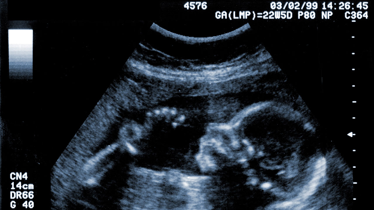
Congratulations on reaching the 20th week of your pregnancy! This is an exciting milestone, as it’s typically around this time that you’ll have your anatomy scan, also known as the 20-week ultrasound. This ultrasound is a detailed examination of your baby’s anatomy to assess their growth and development.
What to Expect During Your 20-Week Ultrasound
Your 20-week ultrasound will typically take place between 18 and 22 weeks of pregnancy. It’s usually performed transabdominally, meaning the ultrasound transducer will be placed on your abdomen to capture images of your baby.
During the ultrasound, your healthcare provider will assess the following:
- Baby’s size and growth: They will measure your baby’s head circumference, abdominal circumference, and femur length to ensure they are growing at an appropriate rate.
- Baby’s anatomy: They will examine your baby’s head, face, neck, chest, abdomen, arms, legs, hands, and feet to check for any abnormalities or birth defects.
- Placenta: They will assess the location, size, and appearance of the placenta, which provides oxygen and nutrients to your baby.
- Amniotic fluid: They will measure the amount of amniotic fluid surrounding your baby, which is important for their growth and development.
- Cervix: They will check the length and condition of your cervix, which is the opening to your uterus.
What the Ultrasound Can Reveal
The 20-week ultrasound can provide valuable information about your baby’s health and well-being. It can help detect:
- Major birth defects: Such as spina bifida, heart defects, and cleft lip or palate.
- Growth abnormalities: Such as intrauterine growth restriction (IUGR) or macrosomia (excessive fetal growth).
- Placental problems: Such as placenta previa (placenta covering the cervix) or placental abruption (placenta separating from the uterine wall).
- Amniotic fluid abnormalities: Such as polyhydramnios (too much amniotic fluid) or oligohydramnios (too little amniotic fluid).
- Cervical issues: Such as cervical shortening or dilation, which may increase the risk of preterm birth.
Preparing for Your 20-Week Ultrasound
To prepare for your 20-week ultrasound, you may be asked to:
- Drink plenty of fluids before the appointment to fill your bladder, which helps improve the ultrasound images.
- Wear loose, comfortable clothing that allows easy access to your abdomen.
- Bring any relevant medical records or test results to the appointment.
After Your 20-Week Ultrasound
After your 20-week ultrasound, your healthcare provider will discuss the results with you and answer any questions you may have. They will also provide guidance on any follow-up care or testing that may be necessary.
If the ultrasound reveals any abnormalities or concerns, your healthcare provider may recommend additional testing, such as:
- Fetal echocardiogram: A detailed ultrasound of your baby’s heart to assess its structure and function.
- Amniocentesis: A procedure to collect amniotic fluid to test for genetic abnormalities or infections.
- Chorionic villus sampling (CVS): A procedure to collect a sample of placental tissue to test for genetic abnormalities.
Conclusion
The 20-week ultrasound is an important milestone in your pregnancy, providing valuable information about your baby’s health and development. By understanding what to expect during the ultrasound and preparing accordingly, you can help ensure a smooth and informative experience. Remember to discuss any concerns or questions with your healthcare provider before and after the ultrasound.