8 Week Pregnancy Images: A Visual Journey of Fetal Development
Pregnancy is a remarkable journey that involves the growth and development of a new life within the mother’s body. At 8 weeks, the embryo has transitioned into a fetus, and significant changes are taking place. Ultrasound images provide a fascinating glimpse into this early stage of pregnancy, revealing the intricate details of the developing fetus.
What to Expect in an 8-Week Ultrasound
An 8-week ultrasound is typically performed transvaginally, using a wand-like device inserted into the vagina. This provides a clearer view of the uterus and its contents compared to an abdominal ultrasound.
During the ultrasound, the technician will use sound waves to create images of the fetus and surrounding structures. These images can reveal:
- Gestational sac: A fluid-filled sac that surrounds the fetus.
- Yolk sac: A small sac attached to the fetus that provides nutrients.
- Embryonic pole: A small, bean-shaped structure that represents the developing fetus.
- Heart flicker: A tiny, flickering image that indicates the presence of a heartbeat.
Fetal Development at 8 Weeks
At 8 weeks, the fetus is approximately 1.5 centimeters (0.6 inches) long and weighs about 1 gram (0.035 ounces). The fetus is rapidly developing, with major organs and structures beginning to form.
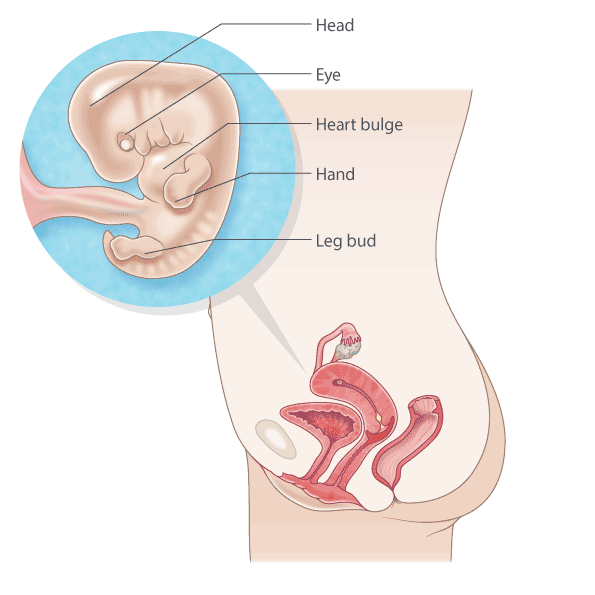
- Head and face: The head is disproportionately large compared to the body, and the facial features are starting to take shape. The eyes, nose, and mouth are visible as small indentations.
- Limbs: The arms and legs are developing, and the fingers and toes are beginning to form.
- Internal organs: The heart, lungs, liver, and kidneys are all present and functioning.
- Nervous system: The brain and spinal cord are developing rapidly.
- Circulatory system: The blood vessels are forming and the heart is pumping blood throughout the body.
Ultrasound Images of 8-Week Fetus
Ultrasound images of an 8-week fetus can vary slightly depending on the equipment used and the position of the fetus. However, some common features include:
- Gestational sac: A dark, circular or oval shape surrounding the fetus.
- Yolk sac: A small, round or oval sac attached to the fetus.
- Embryonic pole: A small, bean-shaped structure located within the gestational sac.
- Heart flicker: A tiny, flickering image within the embryonic pole.
- Limb buds: Small, protruding structures representing the developing arms and legs.
Importance of 8-Week Ultrasound
An 8-week ultrasound is an important milestone in prenatal care. It allows the healthcare provider to:
- Confirm pregnancy: The presence of a gestational sac and embryonic pole confirms the presence of a viable pregnancy.
- Determine gestational age: The size of the gestational sac and embryonic pole can help estimate the gestational age of the fetus.
- Detect multiple pregnancies: An ultrasound can reveal the presence of more than one fetus, such as twins or triplets.
- Identify potential abnormalities: While rare, an ultrasound can sometimes detect potential abnormalities in fetal development.
Conclusion
8-week pregnancy images provide a captivating glimpse into the early stages of fetal development. These images allow healthcare providers to assess the health and progress of the pregnancy and can provide valuable information to expectant parents. As the pregnancy progresses, subsequent ultrasounds will continue to reveal the remarkable transformation of the fetus into a fully formed newborn.