Pregnancy Ultrasound: Capturing the First Glimpses of Your Unborn Child
Pregnancy is an extraordinary journey filled with anticipation and wonder. As you progress through each trimester, your body undergoes remarkable changes to accommodate the growth and development of your unborn child. One of the most captivating moments during this time is the ultrasound scan, which provides a glimpse into the womb and allows you to witness the miraculous development of your little one.
What is an Ultrasound?
An ultrasound, also known as sonography, is a non-invasive imaging technique that uses high-frequency sound waves to create real-time images of the inside of your body. During a pregnancy ultrasound, a transducer (a small, handheld device) is gently placed on your abdomen or inserted into your vagina. The transducer emits sound waves that bounce off the tissues and organs within your uterus, creating a detailed image on a monitor.
Types of Pregnancy Ultrasounds
There are several types of pregnancy ultrasounds, each performed at different stages of pregnancy and serving specific purposes:
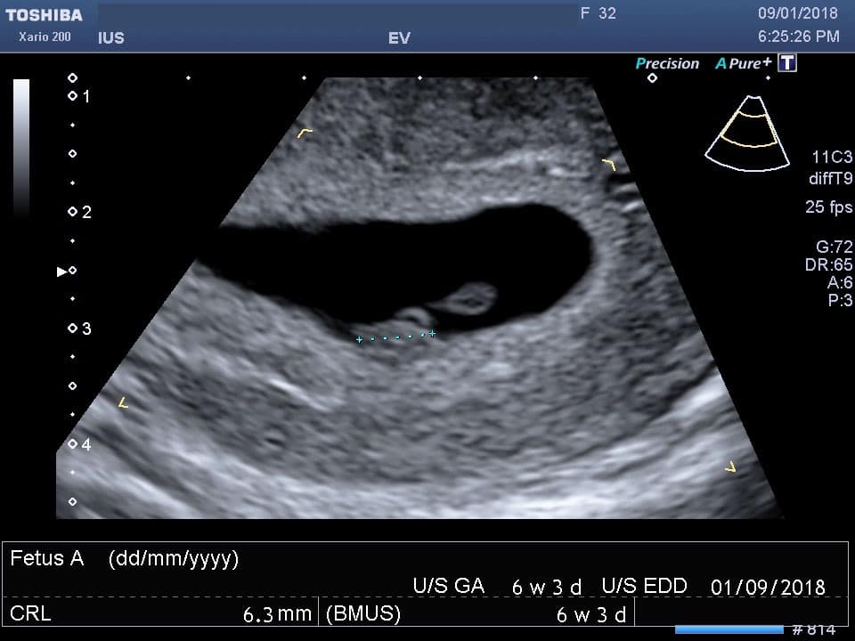
- Transvaginal Ultrasound: This ultrasound is typically performed early in pregnancy (before 12 weeks) and involves inserting a transducer into the vagina. It provides clearer images of the uterus and developing embryo than abdominal ultrasounds.
- Abdominal Ultrasound: This ultrasound is performed later in pregnancy (after 12 weeks) and involves placing the transducer on the abdomen. It provides a broader view of the uterus, placenta, and fetus.
- Nuchal Translucency Ultrasound: This ultrasound is performed between 11 and 14 weeks of pregnancy and measures the thickness of the fluid-filled space at the back of the fetus’s neck. It helps screen for certain chromosomal abnormalities, such as Down syndrome.
- Anomaly Scan: This ultrasound is typically performed between 18 and 22 weeks of pregnancy and provides a detailed examination of the fetus’s anatomy. It helps identify any potential birth defects or growth concerns.
- Growth Scan: This ultrasound is performed in the third trimester and measures the fetus’s growth and development. It also assesses the amount of amniotic fluid and the position of the placenta.
Benefits of Pregnancy Ultrasounds
Pregnancy ultrasounds offer numerous benefits, including:
- Confirming Pregnancy: An ultrasound can confirm pregnancy as early as 5-6 weeks after conception.
- Determining Gestational Age: Ultrasounds can accurately determine the gestational age of the fetus, which helps estimate the due date.
- Monitoring Fetal Growth and Development: Ultrasounds allow doctors to track the growth and development of the fetus throughout pregnancy.
- Identifying Multiple Pregnancies: Ultrasounds can detect multiple pregnancies, such as twins or triplets.
- Screening for Birth Defects: Certain ultrasounds, such as the nuchal translucency ultrasound and anomaly scan, can help screen for potential birth defects.
- Assessing Placental Health: Ultrasounds can evaluate the size, location, and health of the placenta.
- Determining Fetal Position: Ultrasounds can help determine the position of the fetus, which is important for planning delivery.
Safety of Pregnancy Ultrasounds
Pregnancy ultrasounds are generally considered safe and do not pose any known risks to the mother or fetus. The sound waves used in ultrasounds are non-ionizing, meaning they do not contain harmful radiation. However, it is important to note that excessive or prolonged exposure to ultrasound waves should be avoided.
Preparing for an Ultrasound
To prepare for an abdominal ultrasound, you may be asked to drink plenty of water beforehand to fill your bladder. This helps create a clearer image of the uterus and fetus. For a transvaginal ultrasound, you will be asked to empty your bladder before the procedure.
During the Ultrasound
During the ultrasound, you will lie on an examination table. The sonographer will apply a water-based gel to your abdomen or insert the transducer into your vagina. The transducer will be moved around to obtain different views of the uterus and fetus. The entire procedure typically takes around 15-30 minutes.
Interpreting the Ultrasound Results
After the ultrasound, the sonographer will provide you with a report and images of the scan. Your doctor will review the results and discuss any findings with you. If any abnormalities or concerns are identified, your doctor may recommend further testing or monitoring.
Emotional Impact of Pregnancy Ultrasounds
Pregnancy ultrasounds can be an emotionally charged experience for expectant parents. Seeing the tiny heartbeat of their unborn child or witnessing their movements can evoke feelings of joy, awe, and anticipation. Ultrasounds also provide a tangible connection to the pregnancy and help parents bond with their future child.
Conclusion
Pregnancy ultrasounds are an invaluable tool that provides a window into the womb and allows expectant parents to witness the miraculous journey of their unborn child. These scans offer numerous benefits, including confirming pregnancy, monitoring fetal growth and development, screening for birth defects, and assessing placental health. While pregnancy ultrasounds are generally safe, it is important to use them judiciously and follow the guidance of your healthcare provider. By embracing the power of ultrasound technology, expectant parents can gain a deeper understanding of their pregnancy and create lasting memories of their child’s early development.