Pregnancy Ultrasound at 5 Weeks: A Comprehensive Guide
Introduction
A pregnancy ultrasound at 5 weeks is an important milestone in prenatal care. It provides valuable information about the health and development of your pregnancy. This comprehensive guide will delve into the details of a 5-week ultrasound, including what to expect, how to prepare, and what the results may reveal.
What to Expect During a 5-Week Ultrasound
A 5-week ultrasound is typically performed transvaginally, meaning a small wand-like device is inserted into the vagina to obtain images of the uterus and developing embryo. The procedure is generally painless and takes about 15-20 minutes.
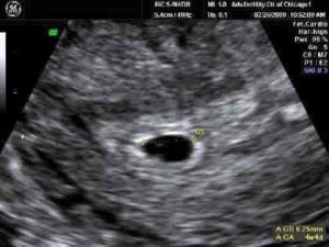
During the ultrasound, the technician will use sound waves to create images of the following:
- Gestational sac: A fluid-filled sac that surrounds the embryo.
- Yolk sac: A small structure that provides nutrients to the embryo.
- Embryo: A tiny, bean-shaped structure that will eventually develop into the baby.
- Fetal heartbeat: A flickering movement that indicates the embryo’s heart is beating.
Preparing for a 5-Week Ultrasound
- Drink plenty of fluids: A full bladder helps push the uterus closer to the front of the body, making it easier to visualize during the ultrasound.
- Avoid gas-producing foods: Foods like beans, broccoli, and cauliflower can cause gas bubbles that may interfere with the ultrasound images.
- Empty your bladder before the procedure: This will help the technician obtain clearer images.
- Bring a support person: Having someone to provide emotional support and ask questions can be helpful.
Interpreting the Results
The results of your 5-week ultrasound will provide important information about the health of your pregnancy.
- Gestational age: The ultrasound will determine how far along you are in your pregnancy, based on the size of the gestational sac and embryo.
- Embryonic development: The ultrasound will assess the development of the embryo, including the presence of a heartbeat and the size of the yolk sac.
- Multiple pregnancies: The ultrasound will reveal if you are carrying twins or more.
- Ectopic pregnancy: An ectopic pregnancy occurs when the embryo implants outside the uterus. The ultrasound can help rule out this possibility.
What if the Results Are Abnormal?
In some cases, the results of a 5-week ultrasound may be abnormal. This does not necessarily mean that there is a problem with your pregnancy, but it may warrant further evaluation.
- No gestational sac: If the gestational sac is not visible, it may be too early to detect or could indicate a miscarriage.
- No fetal heartbeat: The absence of a fetal heartbeat at 5 weeks may be a sign of a miscarriage or a developmental issue.
- Ectopic pregnancy: An ectopic pregnancy can be life-threatening and requires immediate medical attention.
Follow-Up Care
After your 5-week ultrasound, your doctor will discuss the results with you and determine the next steps in your prenatal care. This may include:
- Repeat ultrasound: A follow-up ultrasound may be recommended to confirm the findings or monitor the pregnancy’s progress.
- Blood tests: Blood tests can check for hormone levels and other indicators of pregnancy health.
- Prenatal appointments: Regular prenatal appointments will be scheduled to monitor your pregnancy and ensure the health of both you and your baby.
Conclusion
A pregnancy ultrasound at 5 weeks is a crucial step in prenatal care. It provides valuable information about the health and development of your pregnancy. By understanding what to expect, how to prepare, and what the results may reveal, you can approach this important milestone with confidence and knowledge.