Pregnancy Ultrasound Images: A Comprehensive Guide
Introduction
Pregnancy ultrasound images, also known as sonograms, are an essential tool for monitoring fetal development and ensuring a healthy pregnancy. These images provide valuable insights into the baby’s growth, anatomy, and overall well-being. This comprehensive guide will delve into the different types of pregnancy ultrasound images, their purpose, and how to interpret them.
Types of Pregnancy Ultrasound Images
1. Transvaginal Ultrasound (TVUS)
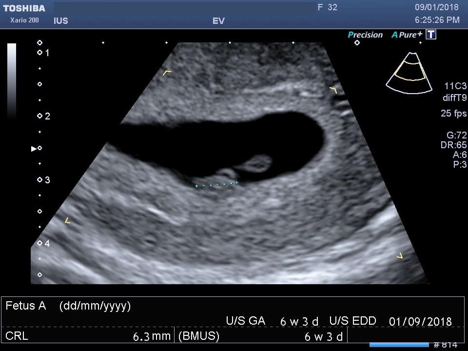
- Performed early in pregnancy (5-12 weeks)
- Uses a probe inserted into the vagina
- Provides clear images of the uterus, ovaries, and early fetal development
2. Transabdominal Ultrasound (TAS)
- Performed later in pregnancy (after 12 weeks)
- Uses a probe placed on the abdomen
- Visualizes the fetus, placenta, and amniotic fluid
3. Doppler Ultrasound
- Measures blood flow in the placenta and umbilical cord
- Assesses fetal well-being and detects potential complications
4. 3D and 4D Ultrasound
- Creates three-dimensional or four-dimensional images of the fetus
- Provides a realistic view of the baby’s facial features and movements
Purpose of Pregnancy Ultrasound Images
1. Confirm Pregnancy
- Detects the presence of a gestational sac and fetal heartbeat
2. Determine Gestational Age
- Measures the fetus’s size and compares it to established growth charts
3. Monitor Fetal Growth and Development
- Tracks the baby’s weight, length, and head circumference
- Assesses organ development and overall well-being
4. Detect Fetal Anomalies
- Identifies structural abnormalities, such as spina bifida or heart defects
5. Evaluate Placental Function
- Determines the placenta’s size, location, and blood flow
- Detects potential complications, such as placental abruption
6. Assess Amniotic Fluid Volume
- Measures the amount of fluid surrounding the fetus
- Indicates potential problems, such as oligohydramnios or polyhydramnios
Interpreting Pregnancy Ultrasound Images
1. Gestational Sac
- Appears as a small, dark circle in early pregnancy
- Gradually increases in size as the pregnancy progresses
2. Fetal Pole
- The first visible sign of the fetus
- Appears as a small, elongated structure
3. Fetal Heartbeat
- Detected as early as 5-6 weeks
- Indicates the presence of a viable pregnancy
4. Nuchal Translucency
- A measurement of the fluid-filled space at the back of the fetal neck
- Used to screen for chromosomal abnormalities
5. Crown-Rump Length (CRL)
- Measured from the top of the fetal head to the bottom of the buttocks
- Used to determine gestational age
6. Femur Length
- Measured from the top of the thigh bone
- Another indicator of fetal growth
7. Placenta
- Appears as a dark, oval-shaped structure
- Attached to the uterine wall
8. Amniotic Fluid
- Surrounds the fetus and appears as a clear, fluid-filled space
9. Umbilical Cord
- Connects the fetus to the placenta
- Visible as a thin, dark line
10. Fetal Movements
- May be observed during later ultrasounds
- Indicate the baby’s well-being
Limitations of Pregnancy Ultrasound Images
- Cannot detect all fetal anomalies
- May be affected by the mother’s body habitus or uterine position
- Does not provide information about the baby’s genetic makeup
Conclusion
Pregnancy ultrasound images are a valuable tool for monitoring fetal development and ensuring a healthy pregnancy. By understanding the different types of images, their purpose, and how to interpret them, expectant parents can gain valuable insights into their baby’s well-being. Regular ultrasound examinations provide peace of mind and allow for early detection of potential complications, leading to optimal outcomes for both mother and baby.