Pregnancy Ultrasound Image: A Comprehensive Guide
Introduction
Pregnancy ultrasound imaging, also known as sonography, is a non-invasive medical procedure that uses high-frequency sound waves to create images of the developing fetus. This technology has revolutionized prenatal care, providing valuable insights into the health and well-being of both the mother and the baby. This comprehensive guide will delve into the intricacies of pregnancy ultrasound images, exploring their significance, interpretation, and potential limitations.
Types of Pregnancy Ultrasound Images
There are several types of pregnancy ultrasound images, each serving a specific purpose:
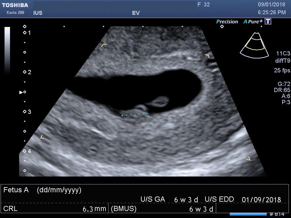
- Transvaginal Ultrasound: This type of ultrasound is performed early in pregnancy, typically between 5 and 12 weeks. A small transducer is inserted into the vagina to obtain clear images of the uterus, ovaries, and developing embryo.
- Transabdominal Ultrasound: This ultrasound is performed later in pregnancy, usually after 12 weeks. A transducer is placed on the abdomen to capture images of the fetus, placenta, and amniotic fluid.
- Doppler Ultrasound: This ultrasound uses sound waves to measure blood flow in the umbilical cord and fetal heart. It can detect potential problems with the baby’s growth and development.
- 3D and 4D Ultrasound: These advanced ultrasound techniques create three-dimensional and four-dimensional images of the fetus, respectively. They provide detailed views of the baby’s facial features, limbs, and movements.
Significance of Pregnancy Ultrasound Images
Pregnancy ultrasound images play a crucial role in prenatal care by:
- Confirming Pregnancy: An ultrasound can confirm pregnancy as early as 5-6 weeks by visualizing the gestational sac and the developing embryo.
- Determining Gestational Age: Ultrasound measurements of the fetus can accurately estimate the gestational age, which is essential for monitoring the baby’s growth and development.
- Evaluating Fetal Health: Ultrasound images can detect potential fetal abnormalities, such as birth defects, growth restrictions, and genetic disorders.
- Assessing Placental Health: Ultrasound can evaluate the size, location, and blood flow of the placenta, which is vital for the baby’s nourishment and oxygen supply.
- Monitoring Amniotic Fluid: Ultrasound can measure the amount of amniotic fluid surrounding the baby, which is important for fetal development and well-being.
Interpretation of Pregnancy Ultrasound Images
Interpreting pregnancy ultrasound images requires specialized training and expertise. Sonographers, who are trained professionals, analyze the images and provide a detailed report to the healthcare provider. The following are key parameters that are assessed during an ultrasound examination:
- Fetal Measurements: These measurements include the head circumference, abdominal circumference, and femur length. They are used to track the baby’s growth and development.
- Fetal Anatomy: The ultrasound images are carefully examined to assess the baby’s organs, limbs, and facial features for any abnormalities.
- Placental Location: The location of the placenta is important to ensure proper blood flow to the fetus.
- Amniotic Fluid Volume: The amount of amniotic fluid is measured to evaluate the baby’s well-being and detect potential complications.
- Fetal Heart Rate: The ultrasound can measure the fetal heart rate, which is an indicator of the baby’s overall health.
Limitations of Pregnancy Ultrasound Images
While pregnancy ultrasound imaging is a valuable tool, it has certain limitations:
- Operator Dependency: The quality and accuracy of ultrasound images depend on the skill and experience of the sonographer.
- Image Resolution: Ultrasound images may not be able to detect all fetal abnormalities, especially those that are very small or located deep within the uterus.
- Time Constraints: Ultrasound examinations are typically limited in duration, which may not allow for a comprehensive assessment of all fetal structures.
- Cost: Ultrasound imaging can be expensive, especially for advanced techniques such as 3D and 4D ultrasounds.
Conclusion
Pregnancy ultrasound imaging is a powerful diagnostic tool that has transformed prenatal care. It provides valuable insights into the health and well-being of both the mother and the baby. By understanding the types, significance, interpretation, and limitations of pregnancy ultrasound images, healthcare providers and expectant parents can make informed decisions regarding prenatal care and pregnancy management.